Get the Most Out of Your CBCT X-ray Machine
Modern dentistry has changed a lot, thanks to 3D dental imaging, especially Cone Beam Computed Tomography (CBCT). Old 2D X-rays were fine, but they squished complex structures into a flat picture. This meant things could look bigger or distorted, and important details might hide behind other structures. It was like trying to see around a corner without moving your head.
CBCT is a big step forward. Unlike regular X-rays that give you a flat view, CBCT X-rays use a cone-shaped X-ray beam. It spins around the patient's head, taking many pictures from different angles. Then, smart software puts these pictures together to create detailed 3D images of the mouth and face. This includes teeth, bone, nerves, and soft tissues. So, you get a full picture without anything overlapping. This helps clear up any guesswork and makes treatment planning much more precise.
This guide is for dental professionals who want to get the most out of their CBCT machine. We want to give you truly useful information. When you understand how to use your CBCT system best—from how to position patients to using advanced software and following safety rules—your practice can get better results, work more smoothly, and, most importantly, build trust with your patients. This guide aims to help you use your CBCT investment to its fullest.
What Your CBCT Can Really Do
 Moving from 2D to 3D imaging isn't just about seeing more; it's about seeing things more clearly and accurately. Old 2D images often had problems like overlapping parts, which made things unclear. CBCT fixes this. It gives you true 3D views, so there's no overlap, and you can measure things precisely from every side. This clearer view means less guesswork, lower risk during complex work, and more predictable treatment results. So, CBCT isn't just a small improvement; it changes how dental professionals diagnose and treat patients.
Here’s a look at what your CBCT can do:
- Implants: It’s vital for planning. You can see the exact bone height, width, density, and important structures like nerves or sinuses before placing an implant. This helps prepare surgical guides, making implant placement safer and more precise.
- Oral & Maxillofacial Surgery: It helps you assess fractures in the face or jaw, pinpoint tumors or cysts, and see exactly where impacted teeth are and if they're near important nerves. It even helps with pre-treatment checks for sleep apnea.
- Orthodontics: You can get a full picture of facial bone structures, how teeth are angled, and assess jaw joint (TMJ) issues. This helps plan complex braces treatments and jaw surgeries.
- Endodontics: It's great for seeing the intricate details of root canals, finding extra canals, spotting root resorption, or precisely locating infections at the tooth's root. High-resolution modes are especially useful here.
- General Dentistry: For everyday care, it helps diagnose complex conditions, find tiny tooth fractures, evaluate dental infections, and clearly assess where impacted teeth are.
Beyond just treatment, your CBCT machine helps you talk to patients better. Those detailed 3D images are great for showing patients exactly what’s going on. When you can show them their own anatomy, the problem, or the planned treatment using detailed 3D models, it really helps them grasp it. This clear, visual explanation empowers patients. They become active, informed partners in their own care. And that builds trust and loyalty, showing how truly helpful content can make your practice a preferred choice.
Getting the Best Image Quality: From Taking the Scan to Reading It
Getting clear, accurate CBCT images starts even before you press the scan button. Proper patient preparation and careful positioning are key. They help avoid image distortions and reduce the need for expensive, time-consuming repeat scans. Even small mistakes in how a patient is aligned can mess up image quality and lead to wrong diagnoses.
Patient Positioning: Getting It Right for Clear Pictures
 Before a patient goes into the scanning area, make sure they take off all removable metal objects from the neck up. This means necklaces, eyeglasses, earrings, dentures, partials, and orthodontic retainers. Doing this stops metal from showing up as artifacts that can hide important details. Also, the person operating the machine needs to pick the right scan setting. This could be high-resolution or low-dose, depending on what the dentist needs to see.
Once ready, the patient should stand as straight as possible, holding the equipment handles firmly. This keeps them steady. Their chin should rest comfortably, and their occlusal plane (the imaginary line between upper and lower teeth) should be parallel to the floor. Most modern CBCT machines have laser guides. These are super helpful for precise alignment. Before the full scan, take a "scout image" or preview scan. This is a low-dose image that lets the operator check and make any small adjustments. Finally, give clear instructions: tell the patient to stay still, breathe normally, and avoid swallowing or moving during the scan.
Proper patient positioning is really important for clear images and avoiding repeat scans. If a patient isn't positioned right, it's much more likely you'll get image artifacts. Retakes expose the patient to extra radiation, take up valuable chair time, mess up the office schedule, and put extra wear and tear on the CBCT machine. So, taking the time to position the patient carefully is an investment in how smoothly your practice runs, patient safety, and how long your equipment lasts.
Understanding Image Problems: How to Spot and Fix Artifacts
Even with careful patient positioning, CBCT images can still have problems like streaks, blurriness, or less contrast. These are called artifacts, and they can make the image quality worse. Knowing what these problems look like and how to fix them is key.
- Metal Artifacts: These appear as streaks, dark bands, or missing information around dense metal objects like fillings or crowns.
- Fix: Remove removable metal. If possible, angle the machine to avoid metal. Use special Metal Artifact Reduction (MAR) software if your system has it.
- Motion Artifacts: These look like blurry images, double outlines, or distorted anatomy. They are caused by patient movement during the scan.
- Fix: Use positioning aids. Give clear patient instructions to stay still. Use the shortest scan time possible.
- Beam Hardening/Scatter: These cause streaks, cupping, or less contrast. They happen when X-rays are absorbed differently and scattered radiation.
- Fix: Adjust kVp settings or use extra beam filters (like copper). Advanced reconstruction software and AI can also help reduce these.
Artifact Type | Appearance/Cause | Operator-Led Mitigation Strategies |
Metal Artifacts | Streaks, dark bands, less contrast, hiding structures; caused by dense metal (fillings, prosthetics). | Remove removable objects; angle the machine to avoid metal; increase kVp; use thin sections; use special Metal Artifact Reduction (MAR) software. |
Motion Artifacts | Double outlines, blurry images, poor overall quality; caused by patient movement during the scan. | Use positioning aids; consider sedation for uncooperative patients; use the shortest scan time possible; tell patients to hold their breath (for chest scans); use "body scan mode" if available. |
Beam Hardening/Scatter | Streaks, cupping, less contrast; caused by X-rays being absorbed differently and scattered radiation. | Adjust kVp settings; use extra beam filters (like copper); use advanced reconstruction software and new AI methods. |
New technologies, especially deep learning (a type of AI), are making images much clearer by reducing motion and other artifacts. These aren't just about clearer pictures. They're about making your work smoother, reducing the need for manual image adjustments, and boosting confidence in reading CBCT images.
Adjusting Scan Settings: Balancing Detail and Safety
 The technical settings you choose for a CBCT scan are important. They affect both the image quality and, very importantly, the patient's radiation dose. Knowing how to adjust these settings helps you follow the ALARA principle (As Low As Reasonably Achievable). This means getting the best diagnostic detail with the least amount of radiation exposure.
Different settings—like Field of View (FOV), mAs (milliampere-seconds), kVp (kilovoltage peak), and resolution (voxel size)—all affect both the patient's radiation dose and the image quality. For example, higher resolution often means a higher dose. The "best" image quality isn't always the highest resolution or the one with the highest dose. Instead, it's the lowest dose that gives you enough information to answer the specific clinical question.
- Field of View (FOV): This shows how much anatomy is covered. A smaller FOV means less radiation dose and often more detail in that specific area. Always use the smallest FOV that covers only the area you need to see.
- mAs: This directly affects the radiation dose and how "noisy" the image is. Use the lowest mAs setting that still gives you a good enough image for your diagnosis. Many modern CBCT units use pulsed X-ray sources to avoid unnecessary exposure.
- kVp: This affects how well X-rays go through tissue and the image's contrast. Higher kVp, with proper beam filters, can sometimes lower the patient's dose while maintaining or improving image quality.
- Resolution (Voxel Size): A smaller voxel size gives you more spatial detail. However, higher resolution settings naturally increase the radiation dose. Only use high-resolution modes when extreme detail is absolutely necessary.
FOV Size | Coverage Area | Typical Use Cases | Radiation Impact | Image Detail |
Small (4x4 cm) | 1–3 teeth | Endo, root fractures, single implant | Low | High |
Medium (8x8 cm) | Quadrant, anterior / posterior arch | Implants, ortho, TMJ | Moderate | Moderate |
Large (16x13 cm+) | Full jaw or craniofacial | Full arch implant, ortho, airway | Higher | Lower |
Leveraging Your Software: Unlocking Advanced Diagnostic Capabilities
Your CBCT machine is just one part of the system. Its viewing software is just as important. This software helps professionals diagnose and plan treatments with great accuracy by making sense of all the data the scanner creates. Good software lets you easily manage different 3D image types, like raw CBCT images, surface models (from intraoral scanners), and 3D photos, all in one place.
Here are some core features of CBCT software:
- Image Viewing & Manipulation: Easily see images from different angles (axial, coronal, sagittal). You can also turn 3D data into familiar panoramic images and cross-sectional slices.
- Measurements & Annotations: Precisely measure lengths, angles, and volumes. Trace nerve canals for safe and accurate planning.
- 3D Rendering View: Get an immediate, full picture of a patient's anatomy. This is also excellent for patient education.
- Superimposition: Layer two CBCT images, or combine CBCT images with intraoral scans or 3D photos. This is powerful for comparing "before and after" views or tracking progress.
- Specialty-Specific Tools: Look for features made for different dental fields, like those for orthodontics, implantology, endodontics, or TMJ analysis.
The Power of AI in Your Software
Adding Artificial Intelligence (AI) to CBCT viewing software is a big step forward. AI isn't meant to replace the dentist. Instead, it helps them do their job better and makes their work smoother, especially with the huge amount of data CBCT creates.
CBCT scans create a lot of data—thousands of image slices. Manually reviewing all this can be slow and even lead to burnout in busy practices. AI helps with this by automating routine tasks, letting you focus more on diagnosis and patient care.
AI-enhanced features include:
- Automated Segmentation and Recognition: AI can automatically find and label different anatomical structures like teeth, nerves, jaws, airways, and sinuses. This speeds up the initial review.
- Virtual Patient Creation: AI can combine CBCT images with intraoral scans and 3D photos. This lets you create a full 3D "virtual patient" for comprehensive treatment planning and patient communication.
- Smart Navigation: Features like automatic tooth number recognition let you easily jump to specific teeth in the scan.
- Efficient Airways Analysis: AI can quickly separate airways, measure their volume, and show their smallest area. This is helpful for conditions like obstructive sleep apnea.
Keeping Your Machine Running: Maintenance, Calibration, and Safety
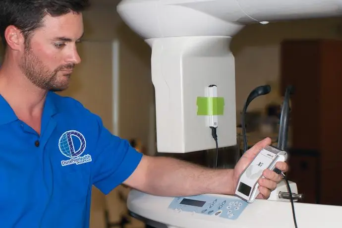 Your CBCT X-ray machine is a big investment. To keep it working well, producing good images, and lasting a long time, you need consistent, regular maintenance and proper calibration.
Routine Maintenance & Calibration: Protecting Your Investment
- Cleaning: After each patient, thoroughly clean and disinfect all parts the patient touched (chin rest, head supports).
- Clear Operating Area: Keep the space around your unit free of clutter so it can spin freely.
- Firmware Updates: Regularly update your unit's software to ensure peak performance.
- Report Issues Quickly: Don't ignore small problems. Report any unusual behavior to an authorized service provider right away.
- Proper Patient Positioning: This is also a maintenance step! It reduces the need for retakes, which lowers radiation exposure and reduces unnecessary wear and tear on the machine.
Calibration is just as important:
- User Calibration: Many systems let you do your own calibration. Aim for at least twice a year, ideally four times. This helps keep image quality consistent.
- Authorized Service: Have a certified technician do a full inspection and calibration at least once a year. This yearly check-up covers all critical components and ensures proper function.
The ALARA Principle in Practice: Minimizing Radiation Exposure
") In dental X-rays, ALARA (As Low As Reasonably Achievable) is the most important rule for radiation safety. It means dentists should keep radiation exposure as low as possible while getting the diagnostic information they need.
In dental X-rays, ALARA (As Low As Reasonably Achievable) is the most important rule for radiation safety. It means dentists should keep radiation exposure as low as possible while getting the diagnostic information they need.
- Justification: Only order a CBCT scan if it's truly needed and other imaging methods can't provide the answer.
- Optimize Scan Parameters: As discussed, carefully choose the FOV, mAs, kVp, and resolution to get diagnostic quality with minimal dose.
- Patient Protection: Use protective thyroid collars and aprons, especially for children and women of childbearing age.
- Dose Monitoring: Pay attention to dose displays on your machine and track radiation dose for patients.
Following the Rules: Regulatory Compliance & Shielding
Running a CBCT machine means you have to follow many rules and standards to keep patients and staff safe.
- Equipment Registration: Register your CBCT system with your state's radiation control program.
- Shielding Plans: You'll likely need a detailed shielding plan for your facility, often prepared by a Qualified Medical Physicist.
- Dosimetry Badges: Staff working near the machine who might receive a certain level of exposure should wear dosimetry badges to track their radiation levels.
- Inspections & QA: Expect regular inspections (e.g., yearly) and quality assurance tests to ensure your facility follows safety rules and your equipment works properly.
Growing Your Skills: Training, Learning, and Seamless Workflow
Getting the most out of a CBCT machine isn't just about buying and installing it. It means committing to ongoing learning and fitting it smoothly into your daily practice.
Learning to Read 3D Images
Many dental professionals, especially those used to 2D X-rays, might find reading 3D CBCT images challenging. This often happens because they didn't get much formal training in advanced radiology during dental school. Without special training, dentists might feel unprepared to analyze and report on the detailed anatomy shown by CBCT scans, including structures beyond just teeth.
To help with this, there are generally two levels of training: Level 1 for dentists who order CBCT scans so they can understand the reports they get, and Level 2 for a deeper understanding and the skills needed to justify, perform, and interpret the scan. Consistent practice, reviewing many different cases, and taking continuing education courses specifically about CBCT scan interpretation are key.
The Need for Ongoing Education
CBCT technology and its software are always changing. So, ongoing professional development is necessary to keep healthcare professionals updated on the latest features, updates, and best practices. This ensures accurate diagnoses, precise planning, efficient use of time, and better patient communication.
Fitting CBCT Smoothly into Your Practice
Bringing new technology like CBCT into a dental practice isn't always easy. You might face:
- Compatibility Issues: New CBCT systems might not work perfectly with your current practice management software.
- Learning Curves: Staff need good training. The time it takes to learn the system can be a big hurdle.
- Managing Lots of Data: A single CBCT scan can have thousands of images. Manually reviewing all this can be overwhelming.
Solutions: Specialized IT support can help with compatibility. AI integration can automate tasks, reducing data overwhelm and making work smoother. Proactive planning for installation and updates also minimizes downtime.
Conclusion: Your CBCT Machine – A Cornerstone of Excellence
 Your CBCT machine is a powerful tool. When you maximize its utility, it transforms how you diagnose and plan treatments, giving you unparalleled insight. It also plays a vital role in building patient engagement and trust, as they can clearly see and understand their own anatomy and treatment plan.
Getting the most out of your CBCT machine isn't just about the technology itself. It’s about a holistic approach: ensuring excellent image quality through proper acquisition, leveraging advanced software features, adhering to strict safety protocols, and committing to continuous learning. By mastering these areas, your CBCT machine becomes a cornerstone of exceptional patient care and a symbol of your practice's commitment to the highest standards, and a significant return on investment (ROI).
Get the Most Out of Your Dental CBCT Machine